"Neural Dedifferentiation" Is One Cause of Cognitive Aging. What Is It, and What Can We Do About It?

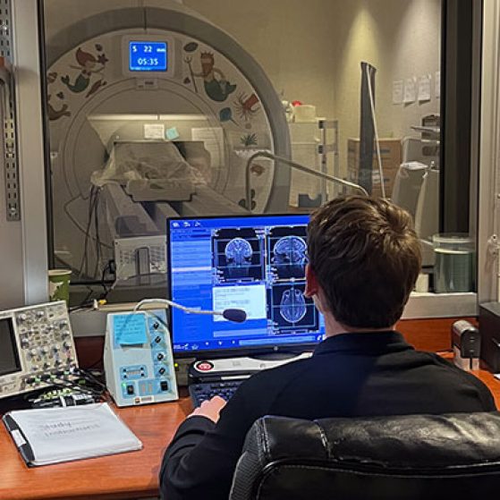
Professor Thad Polk (Cognition and Cognitive Neuroscience) and his lab draw on functional magnetic resonance imaging (fMRI), magnetic resonance spectroscopy (MRS), and other technology to study the physical processes that occur in our brains while we think. Since the early 2000s, Polk’s most-cited body of work has focused on how our brains change as we age. Polk is especially known for his work on neural dedifferentiation, the finding that neural representations become less precise and selective as we get older, which contributes to declines in memory and processing speed that often accompany aging.
To better understand neural dedifferentiation, consider the following scenario: A man—let’s call him Billy Maize—is visiting U-M for our Psychology Homecoming Picnic. But it has been a while since he last visited, and he soon gets lost. As he navigates campus, he searches for landmarks: Burton Tower, the Rackham Building, the Law Quad. As he does so, he also scans for familiar faces. When he nears East Hall, he finally notices two people who seem familiar. He follows them and soon spots a large tent sporting an LSA Psychology banner: he has arrived. Billy enjoys some delicious pulled chicken and mac-and-cheese and has a great time catching up with folks in the department.
If Dr. Polk had been shadowing Billy with a hypothetical portable fMRI machine (the real ones are definitely not portable), he would have observed a few things. As Billy searched for landmarks, Polk would observe a high degree of activity in a region of Billy’s brain known as the parahippocampal place area. In contrast, as Billy looked for people, Polk would see more activity in another region known as the fusiform face area.
But exactly how those processes would play out depends partly on whether Billy is visiting for his 10th Homecoming reunion or his 50th. The younger Billy’s brain would show more selectivity about which neurocognitive circuity was employed for each task. That is, a younger Billy’s brain would show more activity in the place area (and less in the face area) when looking at places and more activity in the face area (and less in the place area) when looking at faces. As Billy ages, however, his brain would show increasing generalization in its responses: fMRI would show more activation of the “incorrect” area when processing either places or faces. In other words, as we age, our neurocognitive responses tend to dedifferentiate: our brains take more of a nonspecific “shotgun approach” to processing varying stimuli.
“Many people (including me!) can relate to the experience of processing information a little more slowly or losing their keys a little more often as they get older,” says Polk. “And our research suggests that neural dedifferentiation might play a role in some of those age-related cognitive impairments. In particular, when we ask a lot of older adults to perform cognitive tasks and then we scan their brains, we find that the people who exhibit the most differentiated or distinctive brain activity patterns also tend to perform the best on a range of cognitive tasks.”
But dedifferentiation does not happen at the same rates for everyone. Many older adults continue to exhibit distinctive brain responses that look like those of their grandchildren, which offers hope that neural dedifferentiation is not inevitable. Given that, it is possible that better understanding dedifferentiation’s causes may eventually allow us to slow or even reverse the process.

To be clear, we are still a very long way from developing a De-Dedifferentiating Elixir of Cognitive Youth. But research by Polk and others has offered some interesting insights. For example, both human and animal studies have shown a positive correlation between levels of the neurotransmitter GABA and “younger-looking” or more selective neuronal activity [1]. Moreover, higher GABA levels in a given region of the brain correlate with more selective activity patterns in that region, but not in other regions without elevated GABA levels. On its own, that research only shows a correlational relationship between GABA and neural selectivity. But other studies using individual animal cells have shown that infusing older cells with GABA causes them to behave in dramatically “younger” ways, while blocking GABA activity in the young cells causes them to behave more like older cells [2]. These studies suggest that declining GABA levels may have a causative role in dedifferentiation.
But even if that causative role exists, simply flooding the brain with GABA will not be the cure for cognitive aging. GABA activity would need to be enhanced in controlled, selective ways in only certain regions of the brain, which is not possible with current medical technology. Moreover, current methods of increasing GABA activity that do exist, such as administering benzodiazepines, come with serious neurocognitive side effects that more than offset any benefit gained from increasing neural selectivity.
Pending further research and future medical technology, Polk insists that the best thing people can do is to focus on things that we do know can help prevent cognitive aging. “The best way to keep your brain healthy as you age is to keep your whole body healthy. Stay active, eat a healthy diet (e.g., a Mediterranean diet), get enough sleep, reduce stress, and stay engaged in positive social environments. Scientific studies have repeatedly demonstrated that following this advice is associated with a healthier brain and more graceful aging,” he says.
So walking around campus and having fun with friends at the homecoming picnic is exactly what the doctor ordered for Billy Maize. But maybe next time stay away from the mac-and-cheese.
References:
1. Chamberlain, J. D., et al. (2021). GABA levels in ventral visual cortex decline with age and are associated with neural distinctiveness. Neurobiology of Aging, Volume 102, 170-177. https://doi.org/10.1016/j.neurobiolaging.2021.02.013.
(https://www.sciencedirect.com/science/article/pii/S0197458021000683)
2. Leventhal, A. G., et al. (2003). GABA and Its Agonists Improved Visual Cortical Function in Senescent Monkeys. Science 300, 812-815. DOI:10.1126/science.1082874
https://www.science.org/doi/10.1126/science.1082874