Michigan News: Scientists CT-scanned thousands of natural history specimens, which you can access for free
Natural history museums have entered a new stage of scientific discovery and accessibility with the completion of open Vertebrate (oVert), a five-year collaborative project among 18 institutions to create 3D reconstructions of vertebrate specimens and make them freely available online.
The project is led by the Florida Museum of Natural History and includes several biologists from the University of Michigan. A paper summarizing the project and its impacts was published online March 6 in the journal BioScience.
Natural history museums got their start in the 16th century as cabinets of curiosity, in which a few wealthy individuals amassed rare and exotic specimens, which they kept mostly to themselves. Since then, museums have become a resource for the public, with exhibits that showcase biodiversity for anyone interested in learning about it.
However, most natural history collections remain behind closed doors, accessible only to scientists who must either travel to see them or ask that a small number of specimens be mailed on loan. The research team behind oVert wants to change that.
“If you require someone to get on a plane and travel to you to collaborate, that’s prohibitive in a lot of ways,” said David Blackburn, principal investigator of the oVert project and curator of herpetology at the Florida Museum of Natural History. “Now we have scientists, teachers, students and artists around the world using these data remotely.”
Between 2017 and 2023, oVert project members took CT scans of more than 13,000 specimens, with representative species across the vertebrate tree of life. This includes more than half the genera of all amphibians, reptiles, fish and mammals.
More than 1,600 specimens from U-M Museum of Zoology collections were scanned as part of the oVert project—mostly mammals, reptiles, amphibians and fish, along with a few birds. The museum also scanned several hundred specimens for partner institutions.
The U-M museum was the third-largest contributor to the project, after the University of Florida and the University of Washington.
“The University of Michigan Museum of Zoology is the home to some of the largest collections of vertebrates in the world. We were able to utilize those collections and our own in-house CT scanner to produce 3D models of thousands of our voucher specimens, as well as material from collaborating collections,” said Cody Thompson, mammal collections manager and associate research scientist at the museum.
The other U-M co-authors of the BioScience paper are Alison Davis Rabosky, Ramon Nagesan, Daniel Rabosky and Gregory Pandelis.
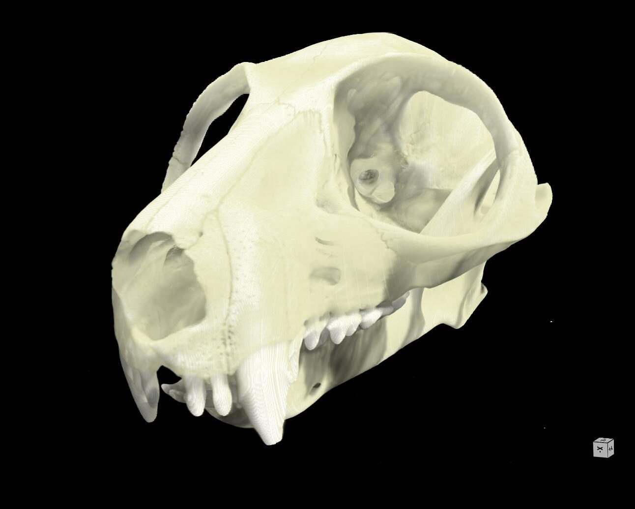
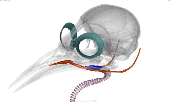
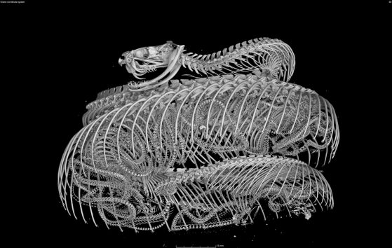
CT scanners use high-energy X-rays to peer past an organism’s exterior and view the dense bone structure beneath. Thus, skeletons make up the majority of oVert reconstructions. A small number of specimens were also stained with a temporary contrast-enhancing solution that allowed researchers to visualize soft tissues, such as skin, muscle and other organs.
The models give an intimate look at internal portions of a specimen that could previously only be observed through destructive dissection and tissue sampling.
“Museums are constantly engaged in a balancing act,” Blackburn said. “You want to protect specimens, but you also want to have people use them. oVert is a way of reducing the wear and tear on samples while also increasing access, and it’s the next logical step in the mission of museum collections.”
oVert was funded with an initial sum of $2.5 million from the National Science Foundation, along with eight additional partnering grants totaling $1.1 million that were used to expand the project’s scope. The goal was to initially scan only specimens preserved in ethyl alcohol, which represent the bulk of fish, reptile and amphibian collections.
Specimens that are too large for fluid preservation are also unable to fit into a CT scanner, but researchers were reluctant to leave these out. A partnering grant to the Idaho Museum of Natural History was used to create a digital model of a humpback whale.
The entire whale was too big to photograph with sufficient resolution, so researchers painstakingly took apart the skeleton, photographed each individual bone, then reassembled the physical and digital specimen.