U-M partners in project to create digital encyclopedia of thousands of 3-D vertebrate specimens
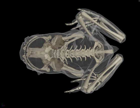
CT scan of Ascaphus truei, the Pacific tailed frog. The “tail” is actually the male genitalia. Images credit: Ed Stanley, Florida Museum of Natural History
A $2.5 million National Science Foundation grant will launch oVert, a new initiative to "teleport" museum specimens from their shelves to the Internet by CT scanning 20,000 vertebrates and making these data-rich, 3-D images available to researchers, educators, students and the public.
The University of Michigan is one of 16 research institutions included in the grant. oVert, short for openVertebrate, complements other NSF-sponsored museum digitization efforts, such as iDigBio, by adding a crucial component that has been difficult to capture—the internal anatomy of specimens.
"The high-resolution, 3-D anatomical data provide the opportunity to compare morphological diversity without damaging specimens. This opens the doors to rare and endangered species that are often difficult to study," said biologist Priscilla Tucker, one of four U-M researchers involved in the project.
U-M is one of five designated scanning centers for the project and will contribute scans from more than 3,500 of its Museum of Zoology specimens—fish, amphibians, reptiles, birds and mammals. CT scanners at the U-M Medical School and School of Dentistry will be used.
With virtual access to specimens through the oVert project, researchers could peel away the skin of a passenger pigeon to glimpse its circulatory system, a class of third-graders could determine a copperhead's last meal, undergraduate students could 3-D print and compare skulls across a range of frog species, and a veterinarian could plan a surgery on a giraffe at a zoo.
The project will encompass representative specimens from more than 80 percent of existing vertebrate genera, and a selection of these will also be scanned with contrast-enhancing stains to characterize soft tissues.
"Each available CT scan provides an opportunity for the community to explore the morphology of a specimen. Those interested in the evolutionary diversity of vertebrates, as well as comparative and functional morphology, can quickly generate new research questions each time they look through high-resolution 3-D anatomical data," said U-M's Tucker, a professor in the Department of Ecology and Evolutionary Biology and curator of mammals at the Museum of Zoology.
U-M was selected as a scanning center for the project because of the size and diversity of the Museum of Zoology's fluid-preserved vertebrate collections, as well as the availability of imaging equipment at the medical and dental schools. The priority will be to scan whole-body, fluid-preserved specimens containing articulated and complete skeletons, Tucker said.
The other U-M researchers involved in the project are Daniel Rabosky, Cody Thompson and Alison Davis Rabosky of the Museum of Zoology and the Department of Ecology and Evolutionary Biology. Rabosky is the lead principal investigator for U-M and Tucker is the co-principal investigator.
Read full Michigan News press release