Nandakumar Lab: Dissecting the telomere–inner nuclear membrane interface formed in meiosis
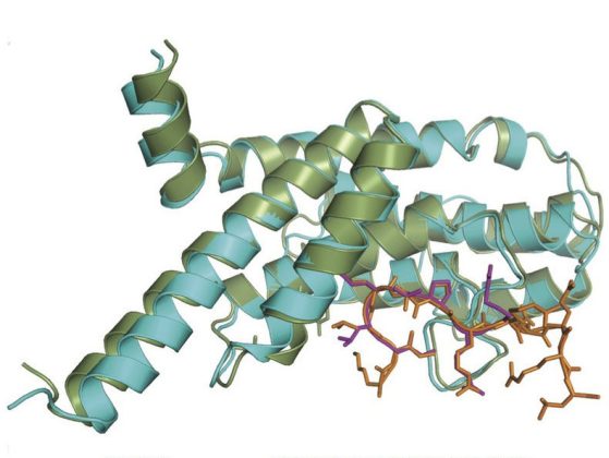
Overlay of the 2 TRF1TRFH-TERB1TRFB complexes: TRFH domains are shown in green and cyan, while the two TBM peptides are shown in stick representation (orange and pink).
A recent study published in Nature Structural & Molecular Biology by JK Nandakumar and his research group used X-ray crystallography to provide a detailed look at the structure of a process called telomere-nuclear membrane interaction that is critical for meiosis, sexual reproduction, and hence, fertility.
Meiosis is a specialized form of cell division that takes parental cells with the usual paired set of chromosomes and produces gametes, or germ cells (egg or sperm) with a single set of chromosomes via one round of DNA replication but two rounds of cell division. The DNA is doubled but then is divided among four daughter cells.
This process contributes greatly to genetic diversity, a process which allows organisms to adapt with changes in their surroundings to survive and continue to propagate. During the first round of cell division in meiosis after DNA replication has already occurred, the chromosomes acquired from each parent of the parent-to-be undergo “genetic crossover”. In this process, fragments of chromosomes are exchanged between the different chromosomes so as to generate a new pair of chromosomes, each of which contain information from both original ones.
Thus when gametes (each of which underwent crossover of chromosomes before meeting the other gamete) from two individuals fuse, the offspring will contain a combination of genetic information from all four grandparents – thus generating great genetic diversity. The importance of meiosis in sexual reproduction is highlighted by the fact that a majority of miscarriages occur as a result of some form of meiotic dysfunction, Nandakumar adds.
For homologous chromosomes to undergo genetic crossover, they have to first physically pair. It has been long known that pairing and crossover depend on the ends of chromosomes, or telomeres, attaching to the nuclear membrane during meiosis. This allows for the chromosomes to move along the nuclear membrane to find their “match”, the homologous chromosome, so as to pair and undergo crossover. However, how telomeres attach to the nuclear membrane and why this occurs only during meiosis was completely unknown until recently.
In 2014, a gene called TERB1 was discovered which expresses only in meiotic cells, and which co-localizes with telomeres and the nuclear membrane during meiosis.
“In the current work we show how TERB1 binds telomeric protein TRF1 to attach telomeres to the nuclear membrane during meiosis,” Nadakumar explains. “We did this by solving the crystal structure of the TERB-TRF1 complex using X-ray crystallography. Our structure shows how TERB1 hijacks the binding strategy used by another telomeric protein (TIN2) to bind TRF1.
"We further show that mutating amino acids at the TRF1-TERB1 interface results in complete loss of formation of this complex. When the same mutations are introduced in mouse meiotic cells (spermatocytes) by our collaborators (Fujiwara et. al), telomeres could no longer attach efficiently to the nuclear membrane for progressing through the rest of meiosis.
“Thus our results provide an atomic picture of telomere-nuclear membrane interaction that is critical for meiosis, sexual reproduction, and hence, fertility,” says Nandakumar. “We predict that mutations in the TRF1-TERB1 interface could account for certain cases of infertility that may exist in the human population."
Link to the paper:
Dissecting the telomere–inner nuclear membrane interface formed in meiosis
Devon F Pendlebury, Yasuhiro Fujiwara, Valerie M Tesmer, Eric M Smith, Hiroki Shibuya, Yoshinori Watanabe & Jayakrishnan Nandakumar
Nature Structural & Molecular Biologydoi:10.1038/nsmb.3493